The chemical engineers at UC Santa Barbara are working on new experiments that are likely to be of great help in detection and diagnosis of pathological tissue related diseases.
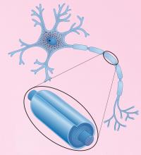 Shown is an illustration of a neuron, which is the basic functional unit of the nervous system. Enlarged portion indicates myelin sheath which is a multilamellar membrane surrounding the axon
The researchers are using nanoscopic imaging methods to study the myelin sheath; this is the membrane that covers the nerves. In people suffering from multiple sclerosis, the thickness of this membrane is reduced.
Jacob Israelachvili, a senior author and a chemical engineering professor at UCSB, explained that the myelin sheath is the communication path for the electric impulses that are transmitted by the central nervous system. He added that sensory and motor disorders are a result of defects in the structure of the myelin membranes. Israelachvili further explained that the onset of disturbances in the membrane in the form of lesions or vacuoles is called demyelination. The scientists observed changes at nanoscopic level by using fluorescence imaging. They also took measurements of the lipid clusters, which are the main components of the myelin membrane, which are most likely to cause lesions. The team studied model molecular layers in various compositions that resemble myelin membranes in healthy and diseased states. They measured the observed differences in size, appearance etc. and created a theoretical model that quantitatively represented the observations. Israelachvili explained that the observed difference between both states is vital for understanding the demyelination process at the single molecule level. According to the research team, the experimental results would be very helpful in the timely detection, diagnosis and treatment of pathological tissue diseases.