A new label-free nanotweezer system uses AI to trap, image, size, and sort milk-derived extracellular vesicles, offering a sharper way to study the nanoscale carriers that could support future drug delivery research.
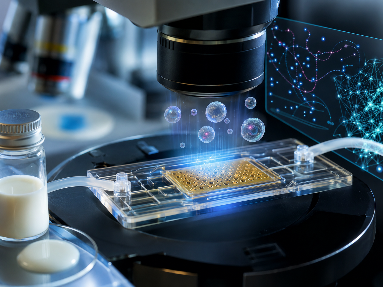
AI-generated illustration based on Lin et al. (2026), Hong, I., Opadele, A.E., Zhu, G. et al. AI-assisted label-free single-particle analysis of milk-derived extracellular vesicles enabled by nanotweezers. npj Biosensing (2026). This image does not reproduce or adapt any original figure from the article.
In a recent research article published in the journal npj Biosensing, researchers developed an advanced electrohydrodynamic nanotweezer platform that enables rapid, label-free trapping and Artificial Intelligence">AI-assisted single-particle analysis of milk-derived extracellular vesicles at the nanoscale.
Nano-Trapping Techniques Overview
Extracellular vesicles (EVs) derived from milk represent a promising platform for drug delivery due to their biocompatibility, ability to traverse the gastrointestinal tract, and potential for immune evasion. However, realizing their therapeutic capabilities requires tools capable of precise, single-particle characterization.
Traditional methods for EV analysis often rely on chemical labeling and ensemble measurements, which can compromise vesicle integrity or obscure heterogeneity. This study introduces a novel nanotechnology platform integrating electrohydrodynamic nanotweezers, interferometric scattering imaging, and artificial intelligence (AI) to enable rapid, label-free, and high-throughput analysis of milk-derived extracellular vesicles (mEVs) at the nanoscale.
Advances in EV Characterization
The core of the platform is an electrohydrodynamic nanotweezer device composed of a thin gold film patterned with an array of micrometer-scale holes, described in the paper as a 15 µm hole array and, in the Results section, as 18 µm microholes with 2 µm lattice spacing.
Application of an alternating current (AC) across this structured gold film induces localized electro-osmotic flow that converges radially toward the microhole centers. This AC electro-osmotic flow effectively traps individual EVs within seconds by counteracting their Brownian motion.
For imaging, label-free interferometric scattering microscopy (iSCAT) is employed, which detects nanoscale particles via interference between incident and scattered light, circumventing the need for fluorescent labels and thereby preserving vesicle integrity.
To automate analysis, a deep learning pipeline based on the U-Net convolutional neural network architecture segments the interferometric images to identify trapped vesicles with high accuracy.
Particle centroids are tracked frame-by-frame, enabling the extraction of Brownian motion trajectories. From these trajectories, diffusion coefficients are calculated to estimate vesicle sizes using the Stokes-Einstein relation. Additionally, by combining Brownian-motion-derived size estimates with simulated contrast curves, the platform inferred the refractive index of each particle, providing insights into purity and heterogeneity without chemical labels.
The study also introduces a frequency-controlled sorting mechanism by varying the AC field frequency to selectively release smaller particles, demonstrating size-based fractionation capabilities of the nanotweezers.
For experimental validation, milk-derived EVs were purified using an acetic acid-based protocol, minimizing protein contamination while preserving EV integrity. Characterization techniques including nanoparticle tracking analysis (NTA), zeta potential measurements, capillary western blotting, and transmission electron microscopy (TEM) complemented the nanotechnology platform to comprehensively assess vesicle properties.
Nanotweezer Platform Insights
The electrohydrodynamic nanotweezer system rapidly trapped thousands of individual mEVs simultaneously, achieving parallel immobilization within seconds. Interferometric imaging successfully visualized label-free vesicles with enhanced contrast following background subtraction.
Using the AI-assisted segmentation and tracking framework, particle trajectories were reconstructed, yielding diffusion-based size estimates with uncertainties quantified via statistical fitting. These size distributions revealed heterogeneous populations primarily in the 150–250 nm range, overlapping with the mid-to-large vesicle fractions detected by NTA, supporting the validity of the nanotweezer platform for the particle populations it interrogated.
Mapping interferometric contrast against size enabled refractive index estimation for individual vesicles, yielding values ranging from 1.38 to 1.50. Particles exceeding this range are likely residual protein aggregates or contaminants, demonstrating the platform’s potential for real-time nanoscale purity assessment. However, NTA also detected smaller subpopulations, including a peak near 43.5 nm, suggesting that the nanotweezer measurements primarily captured the mid- to large-vesicle fractions rather than the full EV size spectrum.
Frequency-dependent manipulation showed that increasing the AC field frequency selectively released smaller particles from traps, effectively performing label-free size sorting. Polymer beads of known sizes validated this size-discrimination capability.
This integrated nanotechnology approach offers several advantages: non-perturbative, real-time analysis that preserves vesicle integrity; scalability through massive parallelization; and enhanced reliability through AI-driven automated processing. By combining electrohydrodynamic trapping with label-free interferometric detection, it addresses several limitations of existing EV analysis approaches, including reliance on labels, ensemble averaging, and limited scalability.
The ability to simultaneously characterize vesicle size and refractive index provides a multifaceted fingerprint that is important for developing EV-based therapeutic systems. Moreover, frequency-controlled release introduces a valuable sorting function, suggesting a route toward future label-free enrichment of EV subpopulations, although downstream biological validation of enriched fractions was not shown.
Implications for EV Therapeutics
This research establishes a powerful nanotweezer-based platform that integrates advanced nanofabrication, electrohydrodynamics, label-free optical detection, and artificial intelligence to advance single-particle analysis of milk-derived extracellular vesicles.
The device harnesses nanoscale fluidic forces generated by AC electro-osmosis on gold microhole arrays to achieve rapid, parallel trapping of vesicles. Combined with interferometric scattering microscopy, this allows real-time, label-free visualization and quantitative assessment of vesicle size and refractive index.
The inclusion of deep learning optimizes detection and tracking for scalable, unbiased characterization. Additionally, frequency-controlled electric fields enable size-selective sorting, potentially supporting sample refinement and downstream applications.
The platform’s nano-scale control and measurement capabilities address critical challenges in EV research by enabling high-throughput, precise, and gentle vesicle analysis. This facilitates a deeper understanding of EV heterogeneity and purity, key factors influencing their biological roles and therapeutic efficacy.
Overall, the study exemplifies how integrating nanotechnology, optics, and AI can provide analytical tools that support the translation of EVs into clinical and biotechnological applications, offering a versatile, scalable platform for future biosensing and drug-delivery research.
Download your PDF copy by clicking here.