A new live-cell AFM method offers scientists a closer look at bacterial membranes, cell poles, and surface changes during growth.
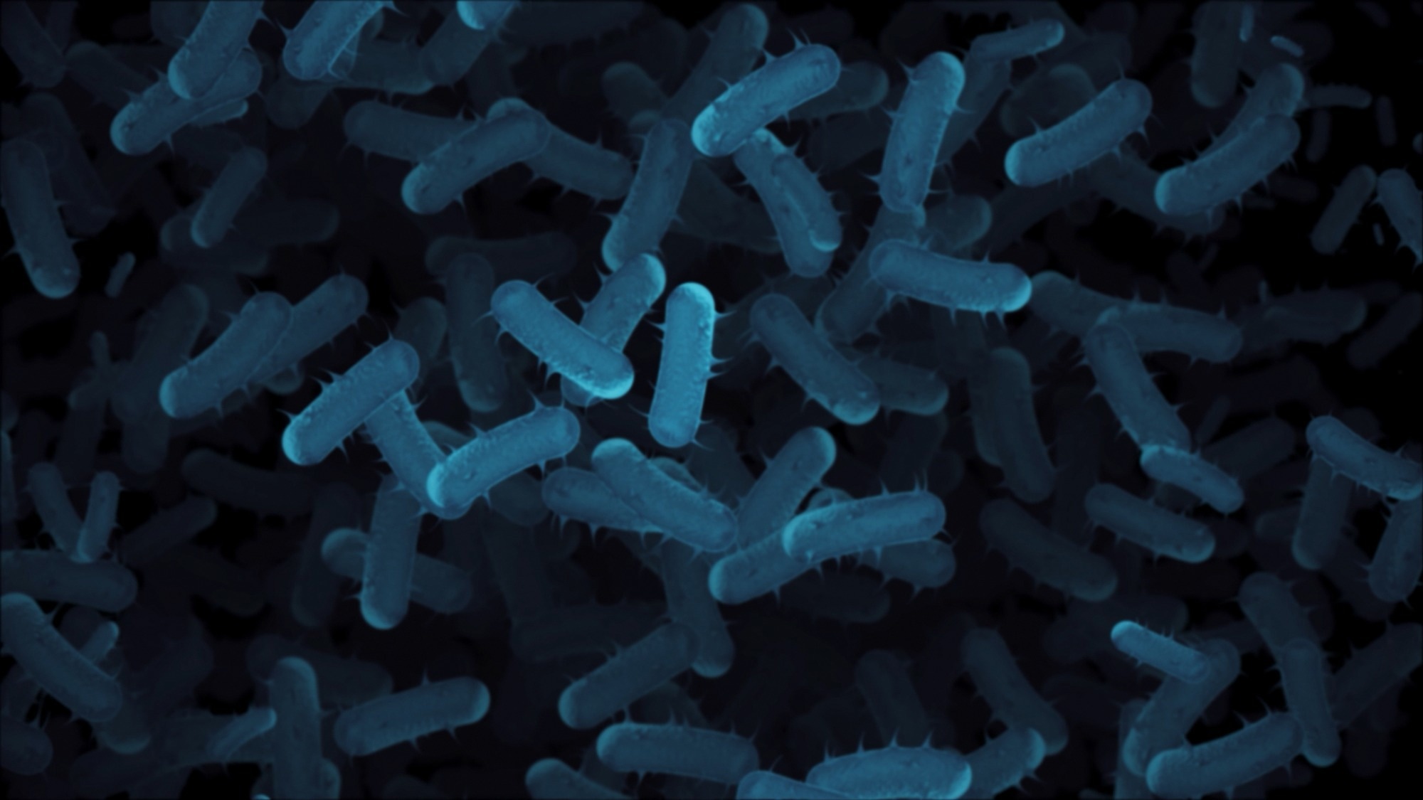
Paper: Nanometer-resolution imaging of living bacteria across species and cellular orientations. Cover Image: Alasabyss / Shutterstock
A recent study published in Nature Communications reports an adapted and refined method for imaging living bacteria at nanometer resolution using atomic force microscopy (AFM).
The researchers adapted a glutaraldehyde-functionalized glass method that enables stable bacterial attachment while preserving high cell viability. In one rough E. coli experiment, attached cells continued to grow and divide. The method allowed detailed comparisons of bacterial outer membrane organization, surface structures, and growth-related changes, and could be used to study antibiotic interactions and bacterial cell biology at the nanoscale.
Addressing the Challenges of Live-Cell Bacterial Imaging
The bacterial cell envelope protects cells from environmental stress and plays a key role in antibiotic resistance. In Gram-negative bacteria, the outer membrane contains lipopolysaccharides (LPS) and membrane proteins that regulate environmental interactions and restrict antibiotic entry. Understanding its molecular organization is important for investigating bacterial defense mechanisms and antimicrobial resistance.
Atomic force microscopy (AFM) enables researchers to examine bacterial surfaces at nanometer resolution, revealing protein networks, peptidoglycan arrangements, and membrane domains.
Live-cell imaging remains difficult because cells must stay firmly attached to the substrate without losing viability. Conventional immobilization methods often provide inconsistent attachment across bacterial species, resulting in cell movement, detachment, and reduced imaging resolution.
The researchers adapted a reversible covalent immobilization method using glutaraldehyde-functionalized glass. The approach enables stable attachment and high-resolution imaging while preserving cell viability. The study also examines how LPS composition, bacterial species, and growth phase influence bacterial envelope architecture.
Developing a Reversible Covalent Immobilization Method
The study compared three surface-functionalization strategies: Vectabond-based electrostatic attachment, glutaraldehyde-mediated covalent attachment, and lectin-based attachment using concanavalin A (ConA). They evaluated these approaches using rough and smooth E. coli strains. Rough strains lack the O-antigen portion of LPS, whereas smooth strains retain this extended polysaccharide structure.
Glass coverslips were cleaned and modified using the respective functionalization procedures. For glutaraldehyde-based immobilization, they first treated the glass with 3-aminopropyltriethoxysilane (APTES) and then applied glutaraldehyde. This process introduced surface-bound aldehyde groups that could react with primary amines on bacterial surfaces.
The reaction forms Schiff base linkages, which provide covalent attachment while remaining reversible in aqueous environments. Protein crosslinking was limited because one aldehyde group was already linked to the glass, and the researchers washed away free glutaraldehyde before adding the bacteria. This helped preserve high cell viability.
Bright-field microscopy quantified bacterial attachment, while membrane-integrity dyes served as a proxy for cell viability. The team then performed AFM imaging to examine bacterial surface structures and evaluate image quality under different immobilization conditions.
The investigation extended to Pseudomonas aeruginosa, Acinetobacter baumannii, and Staphylococcus aureus to test whether the method could support imaging across different bacterial species.
Revealing Bacterial Envelope Architecture at the Nanoscale
The results showed that glutaraldehyde-functionalized surfaces provided more reliable and durable bacterial attachment than conventional electrostatic immobilization.
The improvement was pronounced for smooth E. coli, which attached better to glutaraldehyde-treated glass than to Vectabond-coated surfaces. ConA also attached smooth E. coli efficiently, but it did not hold the cells rigidly enough for high-resolution AFM, and cells frequently detached during imaging.
For rough and smooth E. coli, viability was typically at least 90% over two hours for coatings that successfully retained cells. The researchers also captured a glutaraldehyde-bound rough E. coli cell growing and dividing on the functionalized surface. This showed that at least some immobilized cells could remain active during imaging.
The improved immobilization method enabled a detailed comparison of the outer membranes of rough, smooth, and clear E. coli. Both rough and smooth strains displayed characteristic networks of surface features likely associated with OmpF and OmpC porin trimers. These features appeared as nanoscale depressions distributed across the membrane.
Both strains also contained smooth regions lacking the porin-associated network, supporting earlier identification of these domains as LPS-enriched areas.
The clear E. coli strain, which lacks LPS oligosaccharides, displayed different AFM contrast.
The characteristic porin-associated networks remained largely unresolved, while biochemical analysis confirmed the presence of OmpC and reduced OmpF expression. These findings suggest that polysaccharide chains contribute to the surface contrast that allows AFM to distinguish porin structures. Their absence may change the apparent membrane topography seen by the AFM tip.
The authors also considered increased membrane fluidity, but fixation did not restore the lost contrast, making that explanation less likely.
The method was tested with other bacterial species. Compared with Vectabond, it provided higher or more durable cell attachment in all three tests, including a capsule-free A. baumannii mutant. AFM image quality for A. baumannii appeared similar with both coatings, while S. aureus imaging was more reliable with glutaraldehyde.
The outer membranes of P. aeruginosa and A. baumannii lacked the prominent trimeric porin networks observed in E. coli.
Time-lapse imaging of P. aeruginosa tracked small protrusions of uncertain identity on one cell; their motion lay near the localization noise floor, consistent with a relatively immobile or highly viscous outer membrane. In S. aureus, the researchers observed a mesh-like peptidoglycan cell wall with regions of concentric organization.
The study identified growth-phase-dependent differences in E. coli pole morphology. Stationary-phase cells displayed smooth, rounded poles with recognizable outer membrane protein networks. By comparison, exponentially growing cells frequently exhibited morphological irregularities, and their characteristic protein features remained unresolved. These differences may reflect cell-envelope restructuring and biosynthesis during or soon after cell division.
Expanding the Scope of Live-Cell Nanoscale Imaging
The study shows that glutaraldehyde-functionalized glass can support high-resolution AFM imaging of living bacteria. The method improves attachment when conventional coatings perform poorly. It also extends nanoscale imaging to different bacterial species, strains, and cellular orientations, including poles that are normally difficult to access when rod-shaped cells lie flat on the substrate.
These capabilities support studies of bacterial envelope architecture, membrane organization, and structural changes during cell growth and division. They could also support future studies of antibiotic-bacteria interactions through direct imaging of membrane disruption and cell wall degradation.
Coating conditions may need further refinement for different organisms and experiments, and an immobilization strategy may favor some cells over others, so the sampled cells may not capture the full heterogeneity of the population. Successive cell divisions and biofilm formation remain to be tested over longer imaging periods.
The research broadens the range of bacterial species and cell orientations accessible to live-cell AFM at nanometer resolution.
Disclaimer: The views expressed here are those of the author expressed in their private capacity and do not necessarily represent the views of AZoM.com Limited T/A AZoNetwork the owner and operator of this website. This disclaimer forms part of the Terms and conditions of use of this website.
Source:
- Scheeder, A., Szczepaniak, J., et al. (2026). Nanometer-resolution imaging of living bacteria across species and cellular orientations. Nature Communications, 17, 11329. DOI: 10.1038/s41467-026-78975-y, https://doi.org/10.1038/s41467-026-78975-y