Nanomechanical mapping is essential because the mechanical properties of whole living cells are closely linked to various pathological and physiological processes.
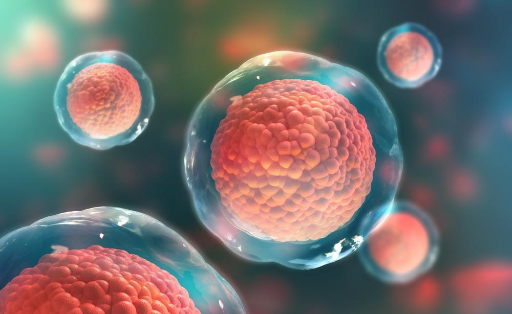
Image Credit: Yurchanka Siarhei/Shutterstock.com
Understanding how changes in the mechanical properties of individual subcellular components or organelles can provide insight into what impacts the cell's overall performance. Scientists have revealed that identifying the factors that lead to the changes in the properties of these components could serve as valuable prognostic markers of diseases.
Nanomechanical Properties of Living Cells
Scientists measure the softness, finite-thickness, viscoelasticity, and incompressibility of living cells via various techniques. Some mammalian cells exhibit elastic modulus values below the 1 kPa range. Studies have shown that eukaryotic cells are softer than a single protein.
Living eukaryotic cells are considered a solid system composed of the plasma membrane enclosing many solid elements, such as protein filaments, DNA, organelles, and a nucleus immersed in an aqueous solution.
Viscoelastic interactions occur when solid elements move in a liquid matrix. Scientists indicated that viscoelasticity is an intrinsic feature of the mechanical response of a cell.
Atomic Force Microscopy (AFM) based measurements describe mechanical properties in terms of the viscoelastic model.
A single cell has been described as anisotropic, heterogenous with finite thickness. Nanomechanical mapping exhibits the structural complexity of a biological component.
The concept of incompressibility of cells has been confirmed by studying the mass densities of HeLa cells and fibroblasts, which were found to be almost equivalent to the water mass density.
Nanomechanical maps characterize the performance of biological molecules. These maps revealed that the local spin of a molecule in thin films could be determined based on the temperature dependence of Young's modulus, from the low to high spin states.
Nanomechanical maps developed by bimodal AFM revealed that the physical properties of lipid bilayers could be modulated by changing the concentration of cholesterol.
Scientists reported that areas of low cholesterol molecule concentration are more elastic than regions with a high concentration.
High spatial resolution nanomechanical maps provide data on the adsorption of single alkali cations on lipid bilayers. Researchers revealed that the development of an ionic network decreases the elastic modulus of the lipid bilayer. These maps also characterize the spatial distribution of magnetic nanoparticles absorbed by liposomes.
Nanomechanical Mapping Techniques
Changes in subcellular components (both molecular or structural aspects), such as the nucleus, nucleolus, and cytoskeleton, are potential markers of diseases. Several methods, including optics-based non-invasive Brillouin microscopy, have helped study the mechanical properties of the subcellular components directly inside living plant and mammalian cells at the submicrometer scale.
Researchers have recently developed a multi-harmonic dynamic AFM (dAFM) method to map quantitatively the nanomechanical properties of soft biological samples in a liquid environment at kHz frequencies
The mapping and imaging capabilities of a force microscope have been challenged while studying living cells, especially eukaryotic cells.
Researchers have reported that living eukaryotic cells are amongst the softest materials on earth. To date, the images of these cells are best obtained via atomic force microscopy that contains a spatial resolution in the 100 nm range.
AFM measures the mechanical properties of the cell by measuring the interaction forces with respect to the AFM tip and surface distance or interaction force on some parameters of the tip's oscillation.
Nanomechanical mapping methods are classified with respect to different features, such as quantitative characters or the ratio between the resonant frequency and the modulation frequency.
Bimodal AFM has been extensively used for nanomechanical mapping. For example, the nanomechanical mapping of enucleated cells has helped elucidate the contribution of the nucleus to the passive cell mechanics.
Application of Nanomechanical Mapping
Nanomechanical mapping helps understand the differences in outcomes of microsurgeries in eyes.
Studies have revealed that many ocular diseases are related to changes in the mechanical and material properties of the eye. Some of the ocular diseases associated with mechanical alterations include macular holes and pucker.
These diseases occur due to abnormal physical tractions acting on the eye retina, and can be corrected via the microsurgical procedure, which includes mechanical peeling of the internal limiting membrane of the retina (ILM).
Scientists performed a nanoscale elastic measurement to understand the differences in the biomechanical response of the ILM in the two diseases, which is also associated with differential outcomes after microsurgeries.
Nanomechanical mapping of the retina helps ophthalmologists develop microsurgical protocols based on the material ILM properties in macular hole or pucker.
A study of protein nanomechanics help scientists understand how mechanical force shapes the free energy of proteins. Researchers studied how nanomechanics is controlled by several factors that are present in living tissues or by mutations.
Identification of specific factors could serve as a target, and it would immensely benefit the development of drugs for the disease.
High-spatial resolution nanomechanical maps characterize the elastic properties of single proteins, membrane proteins, protein fibrils, and DNA. Scientists have also generated nanomechanical maps for bones and virus-like particles.
Continue reading: Using Nanotechnology to Investigate Neural Interfaces
References and Future Reading
Efremov, Y.M., Suter, D.M., Timashev, P.S. et al. (2022) 3D nanomechanical mapping of subcellular and sub-nuclear structures of living cells by multi-harmonic AFM with long-tip microcantilevers. Sci Rep 12, 529 https://doi.org/10.1038/s41598-021-04443-w
Gisbert, G.V. et al. (2021) High-Speed Nanomechanical Mapping of the Early Stages of Collagen Growth by Bimodal Force Microscopy. ACS Nano. 15(1). pp. 1850–1857. https://doi.org/10.1021/acsnano.0c10159
Alegre-Cebollada, J. (2021) Protein nanomechanics in biological context. Biophysical Reviews. 13. pp. 435–454. https://doi.org/10.1007/s12551-021-00822-9
Efremov, M. Y. et al. (2020) Nanomechanical properties of enucleated cells: contribution of the nucleus to the passive cell mechanics. Journal of Nanobiotechnology.18(134). https://doi.org/10.1186/s12951-020-00696-1
Stühn, L. et al. (2019). Nanomechanical sub-surface mapping of living biological cells by force microscopy. Nanoscale. 11. https://pubs.rsc.org/en/content/articlelanding/2019/nr/c9nr03497h
Efremov, Y.M., Cartagena-Rivera, A.X., Athamneh, A.I.M. et al. (2018) Mapping heterogeneity of cellular mechanics by multi-harmonic atomic force microscopy. Nat Protoc 13, 2200–2216 (2018). https://doi.org/10.1038/s41596-018-0031-8
Disclaimer: The views expressed here are those of the author expressed in their private capacity and do not necessarily represent the views of AZoM.com Limited T/A AZoNetwork the owner and operator of this website. This disclaimer forms part of the Terms and conditions of use of this website.