Dec 16 2013
From a heart-shaped cell nucleus to a 3D molecular syringe, creative scientists at the University of Bristol have revealed the beauty found in complex and technical research.
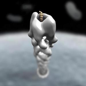
 ‘Staring Down the Barrel of a Gun’, by Martin Cheung
The University of Bristol’s annual Art of Science Competition challenged researchers to look for aesthetic beauty in their laboratories to help make their work more accessible to the public.
There were over 63 entries this year, capturing a range of intriguing and eye-catching subjects from slices of live brain tissue, a DNA helix made from DNA and a microscopic fluorescent image of a fruitfly’s circulatory system.
The 12 winning shots will be on display in the At-Bristol cafe from the end of January and are currently being made into a 2014 calendar.
Among the winners was Rachel Curnock, from the School of Biochemistry, who captured a striking image of a HeLa cell - an immortal cell line used in scientific research – which had an unusual heart-shaped nucleus.
Another intriguing entry was from Martin Cheung, in the School of Cellular and Molecular Medicine, who used electron microscopy to reconstruct a molecular syringe that many pathogenic bacteria use to cause disease.
Becky Brooks, a PhD student in the School of Biochemistry and co-ordinator of the competition, said: “The Art of Science competition is a great opportunity for scientists to show their creative sides, and the entries certainly didn't disappoint.
“The images have already sparked curiosity amongst fellow scientists about the work people do here at Bristol and I hope that this sort of thing will make science accessible to non-scientists too.”
Many of the winning entries will be selected and submitted to Wellcome Images - a biomedical collection which holds over 40,000 high-quality images from the clinical and biomedical sciences, and is the world's leading source of images of medicine and its history.
The submissions were judged and selected according to their scientific and artistic content by Professor George Banting, Dean of the Faculty of Medical and Veterinary Sciences, and Sabrina Taner, Biomedical Image Acquisitions Coordinator at Wellcome Images.