From drug-carrying nanoparticles to ROS-scavenging nanozymes and ultrathin radiation shields, researchers are exploring how materials engineered at the nanoscale could address multiple major pathways of radiation injury simultaneously.
Paper: Protective mechanisms and therapeutic promise of nanomaterials in radiation-induced diseases
Radiotherapy, a key approach in treating cancer, can damage surrounding healthy tissues because ionizing radiation cannot be confined exclusively to cancer cells. Nanomaterials have recently gained attention for radioprotection, as they can provide novel pathways to alleviate or prevent these side effects.
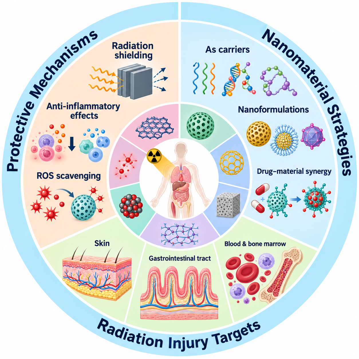
A paper recently published in the International Journal of Pharmaceutics: X provided a comprehensive review of recent developments in radioprotective strategies based on nanomaterials. The review identified several complementary strategies, including delivering protective drugs, suppressing inflammation and oxidative stress, and physically shielding tissues from radiation.
Nanomaterials Delivering Synthetic Drugs
In one study, Wang et al. (2024) leveraged calcium ions' ability to bind with amifostine (AMF) and synthesized AMF-loaded calcium carbonate cores (CC/AMF) to optimize oral delivery. A phase-transfer co-precipitation method was used to prepare CC/AMF.
Later, hybrid nanoparticles (LCC/AMF NPs) were obtained by modifying the core using phospholipids. The hybrid material combined the oral-delivery benefits of lipid NPs with the enhanced drug-loading capacity of calcium carbonate.
It enabled efficient intestinal absorption and transport, thereby significantly enhancing the AMF’s radioprotective effect in vivo via oral dosing.
Similarly, Zhao et al. (2023) synthesized a drug delivery system by first conjugating a PEGylated phospholipid to AMF and then self-assembling the resulting sample with polysorbate 80 and poly(lactide-co-glycolic acid).
This system was capable of alleviating radiation-induced glial cell activation and neuronal damage and penetrating the blood-brain barrier, offering a suitable approach for brain radioprotection.
In another study, Liu et al. (2021) developed a reactive oxygen species (ROS)-responsive drug carrier to co-load WR-1065 and curcumin by conjugating curcumin to poly(ethylene glycol)-poly(ε-caprolactone) through a thioketal linker.
The functional nanodrug was efficiently absorbed by cells, reduced the metabolism of WR-1065 and curcumin within the gastrointestinal tract, and was distributed to several organs. Thus, it protected the hematopoietic system from radiation-induced damage.
Nanomaterials with Anti-inflammatory Effects
El-Ghazaly et al. (2017) found that administering selenium NPs to rats reduced the white blood cell (WBC) count and the levels of total nitrate/nitrite, thiobarbituric acid reactive substance, prostaglandin E2, and tumor necrosis factor-α in paw exudate after radiation induction.
Guo et al. (2024a) introduced oxidized chondroitin sulfate as a polysaccharide-like component of the extracellular matrix in a multifunctional glycopeptide hydrogel, which can accelerate the repair of chronic inflammatory injury by adsorbing inflammatory factors and regulating the inflammatory response.
Kumar et al. (2016) reported that tea polyphenols exerted radioprotective effects by reducing Bax expression and restoring redox balance, thereby mitigating radiation-induced apoptosis and oxidative damage.
The researchers further showed that the bioavailability of tea polyphenols could be improved by encapsulating them using chitosan as the outer shell and bovine serum albumin as the core matrix. Moreover, in a murine model, Jia et al. (2022) found that orally administered polydopamine NPs, a synthetic melanin-like polymer, showed significant potential to scavenge ROS and inhibit inflammatory responses.
In human lymphocytes, Zal et al. (2018) found that cerium dioxide NPs reduced the occurrence of necrosis and apoptosis induced by ionizing radiation. Additionally, cerium dioxide NPs significantly decreased ionizing radiation-induced intracellular IL-1β production.
Shielding Nanomaterials for X-rays
Nano-clays exhibit a radiation-shielding effect that can be used to attenuate X-rays. Sakher et al. (2022) found that green clay from Adrar in the Algerian Sahara demonstrated more effective radiation shielding than Timimoune and Reggan, with radiation attenuation of 99.8% and a mass absorption coefficient of 243.4 cm²/g, respectively.
Composite nanomaterials have also demonstrated X-ray shielding capability. Osman et al. (2023), for instance, integrated lead oxide particles into polystyrene to form polystyrene/lead oxide nanocomposites, whose shielding capabilities were regulated by the lead oxide particle size and concentration.
Similarly, Asadpour et al. (2023) found that including tungsten trioxide and bismuth trioxide nanofillers in a dimethylpolysiloxane matrix enhanced X-ray shielding performance compared with poly- and monometallic fillers.
Kim and Byun (2022) synthesized nanofibers with a multilayer film structure from polymer-tungsten composites using electrospinning. For 60 keV, the resulting nanofiber-based paper demonstrated 64.88% X-ray shielding at 0.1 mm and 90.10% at 0.3 mm.
Zaroushani et al. (2016) fabricated nanocomposites by embedding 15–35 nm nickel oxide nanopowders into EI-403 epoxy resin. Composites containing 7% nickel oxide at 6 mm, 7% at 4 mm, and 11% at 2 mm achieved shielding efficiencies of 64.52%, 66.72%, and 46.80%, respectively.
Nanomaterials for ROS Scavenging
Qiao et al. (2014) found that, at 10 μg/mL, graphene oxide effectively scavenged ROS, decreasing cell death and deoxyribonucleic acid (DNA) damage by 39% and 48% in fibroblasts, respectively, under X-ray exposure.
Xie et al. (2019a) reported that graphene-bovine serum albumin NPs restored the levels of malondialdehyde and superoxide dismutase and improved the scavenging efficacy of radical species. This protected mouse bone marrow DNA and improved cell survival under ionizing radiation.
Xie et al. (2022) used sodium hyaluronate hydrogels loaded with nano-graphdiyne for dermal application to scavenge free radicals. Separately, Zhao et al. (2021) found that fullerenol significantly improved cell viability and blocked ROS-induced damage after radiation in human keratinocytes.
Jahan et al. (2024) incorporated cerium oxide NPs into electrospun poly(ε-caprolactone) (PCL) fibers to reduce cytotoxicity from direct NP exposure while improving cell survival and retaining ROS-scavenging activity.
These findings indicate that the biocompatibility of cerium oxide-based antioxidant systems may be enhanced through polymer encapsulation.
In conclusion, this review highlighted the advances in nanomaterial-mediated radioprotection through shielding, targeted drug delivery, ROS regulation, and anti-inflammatory mechanisms. However, most approaches remain preclinical, and further research is needed to establish long-term safety, biodistribution, scalability, and whether healthy tissues can be protected without reducing tumor control.
Disclaimer: The views expressed here are those of the author expressed in their private capacity and do not necessarily represent the views of AZoM.com Limited T/A AZoNetwork the owner and operator of this website. This disclaimer forms part of the Terms and conditions of use of this website.
Source:
- Ge, Y., Xu, M., Yang, X., Yun, W., Huang, W., & Liu, J. (2026). Protective mechanisms and therapeutic promise of nanomaterials in radiation-induced diseases. International Journal of Pharmaceutics: X, 100658. DOI: 10.1016/j.ijpx.2026.100658, https://www.sciencedirect.com/science/article/pii/S2590156726001787