A switchable DNA structure designed to stay dark until it encounters tumor-associated targets could give surgeons a clearer view of hard-to-reach oral and maxillofacial tumors.
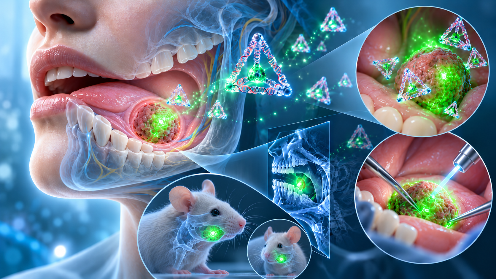
Paper: Fabrication of dynamic tetrahedral DNA structure probe for precision imaging and surgical navigation of oral and maxillofacial tumors. AI-generated abstract conceptual image created using ChatGPT/OpenAI
*Important notice: This news reports on an unedited version of an accepted paper and is awaiting final editing. Therefore, the paper should not be regarded as conclusive or treated as established information.
In a recent research article in the Journal of Nanobiotechnology, researchers created a tetrahedral DNA structure (TDN) probe with a target-responsive fluorescence switch and tested it in cells and mice for precision imaging and fluorescence-guided surgical navigation for oral and maxillofacial tumors.
The findings provide preclinical evidence for a possible imaging and surgical-guidance technique that still requires clinical testing.
Nanoprobe rationale
Oral and maxillofacial tumors can severely affect facial structure and functions such as speaking, breathing, and chewing. Conventional imaging can have limited sensitivity and resolution and can be affected by artifacts. DNA nanoprobe-based imaging, particularly fluorescence in situ hybridization (FISH), offers high specificity and real-time visualization, yet DNA probes can face problems with biological stability and cell affinity.
Tetrahedral DNA nanostructures (TDNs) can address some of these problems. Their defined 3D geometry and biophysical properties, including caveolin-mediated cellular uptake and resistance to nuclease degradation, help maintain biostability and preserve function in physiological environments.
Compared with conventional probes, earlier studies cited by the authors report that TDN-based sensors can provide very high sensitivity, low detection limits, reduced steric hindrance, better-defined probe orientation, and lower background noise, which can improve target accessibility.
The researchers combined TDNs with FISH to create a switchable "on-off" DNA nanoprobe for imaging and surgical navigation of oral and maxillofacial tumors.
TDN-probe design and validation
Researchers designed a switchable "on-off" TDN-probe with a detection strand (S1) containing target-recognition and TDN-hybridizing domains. They placed a fluorescent reporter and a nearby quencher on adjacent parts of the assembled probe. Without a target, fluorescence is quenched. Target binding detaches S1 through strand displacement, restoring fluorescence.
The researchers made the probe by thermal annealing and assessed its structure and target-responsive behavior with gel electrophoresis, atomic force microscopy, transmission electron microscopy, and fluorescence spectroscopy.
For in vitro tests, the researchers tested separate probe configurations targeting nucleolin or microRNA-21 in human melanoma cells (A375), human tongue squamous cell carcinoma cells (CAL27 and SCC9), and mouse fibroblasts (L929) as a non-tumor control.
In mice, the team used axillary and floor-of-mouth, or sublingual, tumor models. The team administered nucleolin-targeting TDN probes via local infiltration or intravenous injection for real-time imaging and fluorescence-guided tumor resection. Resected tissues underwent ex vivo imaging and H&E staining. Fluorescein sodium and non-targeted probes served as controls.
The team followed the animals for two weeks after surgery, assessing wound recovery, behavior, tumor recurrence or metastasis, and the histology of major organs.
Imaging and surgical navigation performance
In vitro experiments showed selective fluorescence associated with nucleolin on cancer cell membranes. Fluorescence intensity was significantly higher in the tumor cell lines A375, CAL27, and SCC9 than in the L929 fibroblast control. Signals reached 23% to 39% of the measured maximum within 1 hour and rose further over 5 hours.
For intracellular imaging, the TDN-probe was modified to target microRNA-21. Strong fluorescence signals appeared within the tumor cells after 12 hours of incubation, while L929 cells showed no significant signal, indicating intracellular target detection.
The lack of significant fluorescence after introducing anti-microRNA-21 to reduce target expression further supported specificity. Intracellular fluorescence recovery was slower than membrane targeting, reaching 43% to 72% of the measured maximum after 4 hours and the measured maximum after 20 hours. The authors linked the slower response to endocytosis.
In vivo studies with superficial axillary tumors and deeper floor-of-mouth tumors also showed the probe's imaging performance. After local infiltration, strong fluorescence appeared at tumor sites within 30 minutes.
Intravenous injection produced visible signals within 10 minutes in the superficial tumor models. In the floor-of-mouth models, signals first appeared at 10 minutes for A375 tumors and 20 minutes for CAL27 tumors. Deeper tumors produced weaker fluorescence than superficial tumors.
Fluorescence-guided surgery with the TDN-probe enabled removal of the fluorescently labeled tumor tissue, with no residual fluorescence detected at the surgical sites. The excised tumors were intact and did not include obvious excess normal tissue.
The authors cautioned that loss of fluorescence does not prove surgical margins are free of tumor cells and called for systematic pathological assessment of the margins in later studies.
Compared with the TDN-probe, fluorescein sodium (FS), used as a positive control, produced intense but non-specific fluorescence and significant residual signals after resection. It was also associated with more extensive tissue removal and greater injury, particularly in the floor-of-mouth model.
Control groups given non-targeted TDN-probes or double-strand DNA probes showed no significant fluorescent signals in tumor regions. The researchers attributed this to the non-targeted probe's failure to trigger strand displacement and to limited tissue penetration and structural instability in the double-strand DNA probe.
The rapid in vivo fluorescence response may partly reflect the TDN-probe's nanoscale size of about 10 nm.
The authors proposed that the enhanced permeability and retention (EPR) effect, in which abnormal tumor blood vessels can allow nanoscale material to enter tumor tissue, may aid tumor accumulation.
They also suggested that the probe's size may support diffusion through dense tumor stroma by reducing steric hindrance. The study did not directly measure the contribution of these proposed mechanisms.
TDN-probe clinical potential
The study created a relatively simple target-responsive TDN nanoprobe and tested it for real-time tumor imaging and surgical navigation in oral and maxillofacial tumor models.
The nanoprobe's ease of synthesis, physicochemical properties, and functional features make it a candidate for further study as a tumor-imaging tool. Its approximately 10 nm size and structural stability may help it remain in and move through tumor tissue, including deeper oral and maxillofacial sites.
The controllable "on-off" response lets the TDN probe produce high-contrast tumor images with lower background fluorescence. Researchers can also change the recognition sequence to detect different tumor biomarkers, as shown by the separate nucleolin- and microRNA-21-targeting versions. The animal imaging and surgical-navigation experiments used the nucleolin-targeting version.
The work remains preclinical. The TDN-probe helped identify labeled tumor tissue for resection in mice without obvious removal of excess adjacent normal tissue. This result suggests that the approach could help surgeons protect nearby maxillofacial structures, but the study did not directly test injury to individual nerves or blood vessels.
No patients were studied. Postoperative follow-up lasted two weeks, and systematic pathology did not confirm the absence of residual fluorescence as a negative surgical margin. The principal in vivo fluorescence analyses reported an n of 3.
Longer studies and larger animal experiments will be needed to assess delayed toxicity, recurrence, margin accuracy, and performance before human testing. Human safety and clinical usefulness remain untested.