A protein-templated iron oxide nanoparticle combines two ROS-scavenging activities with MRI contrast, giving researchers a new way to investigate treatment and disease assessment in the same preclinical platform.
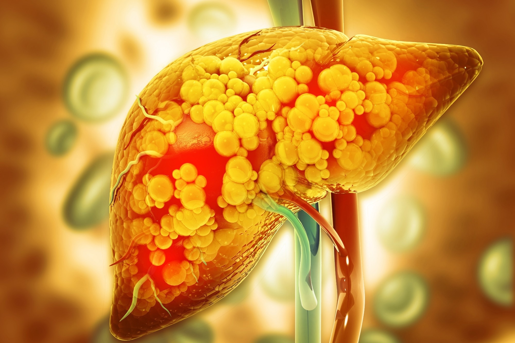
Paper: Biomimetic Cascade Nanozymes: Superoxide Dismutase-Templated Ultrasmall Iron Oxide for Liver Fibrosis Therapy and MRI-Assisted Staging. Image Credit: Crystal Light / Shutterstock
In a recent research article published in the journal Pharmaceutics, researchers developed a biomimetic cascade nanozyme platform that integrates ultrasmall iron oxide nanoparticles templated by natural superoxide dismutase and evaluated it for liver fibrosis therapy and MRI-assisted disease-stage assessment.
Liver Fibrosis and ROS Pathology
Liver fibrosis is a serious condition resulting from chronic liver injuries and characterized by excessive extracellular matrix buildup, primarily collagen, which impairs liver function and may progress to cirrhosis or cancer. Current diagnostic methods, such as liver biopsy, are invasive and limited by sampling error, while noninvasive imaging cannot assess molecular mechanisms.
Therapeutic options to halt or reverse fibrosis remain scarce, and researchers are studying antioxidant therapy because reactive oxygen species (ROS) play a critical role in fibrosis progression by activating hepatic stellate cells to produce collagen. Natural superoxide dismutase (SOD) effectively neutralizes superoxide radicals but faces challenges, including instability and a short circulation time.
Ultrasmall iron oxide nanoparticles can act as magnetic resonance imaging (MRI) contrast agents, and some iron oxide nanomaterials have catalase-like activity that decomposes hydrogen peroxide, a secondary ROS product.
The researchers proposed that a nanosystem integrating both SOD and ultrasmall iron oxide could create a cascade antioxidant system that sequentially removes ROS intermediates while providing MRI contrast for fibrosis assessment.
Synthesis and Characterization of iSOD
The researchers developed a biomimetic nanoplatform named iSOD by using natural SOD as a biological template for a one-pot coprecipitation synthesis of ultrasmall iron oxide (Fe3O4) nanoparticles.
This process yielded spherical nanoparticles with an inorganic core diameter of approximately 4 nm, encapsulated by an active protein shell. Multiple characterization techniques, including transmission electron microscopy and light scattering measurements of particle size, confirmed the ultrasmall core size and colloidal stability.
Enzymatic assays confirmed combined superoxide dismutase and catalase-like activities, showing a dual-enzyme cascade that scavenges ROS by converting superoxide radicals to hydrogen peroxide and then decomposing it into water and oxygen.
In vitro, the antioxidant effect was tested on hepatic stellate cells under oxidative stress. For in vivo evaluation, rat models with varying degrees of liver fibrosis received intravenous iSOD treatment three times weekly over two weeks, with three rats in each treatment subgroup.
MRI was performed before and 30 minutes after injection to compare hepatic signal changes across healthy, moderate fibrosis, and severe fibrosis groups. Histological analyses evaluated collagen deposition and tissue architecture. Short-term safety was assessed separately in healthy mice at days 1 and 7 using blood tests, organ histology, and Prussian blue staining.
Nanozyme Performance and Therapeutics
The iSOD nanozymes contained an ultrasmall iron oxide core of about 4 nm, while the complete protein-coated particles had a hydrodynamic diameter of about 28 nm. The size difference supported successful protein templating, and the particles remained stable in the tested aqueous and physiological media.
Functionally, iSOD demonstrated a sequential enzymatic cascade: the SOD shell scavenged superoxide anions, and the iron oxide core showed catalase-mimetic activity that decomposed the intermediate hydrogen peroxide.
This dual functionality was designed to reduce hydrogen peroxide accumulation associated with SOD activity. The experiments did not directly compare iSOD with free SOD or iron oxide alone. In hepatic stellate cells, iSOD significantly reduced intracellular oxidative stress and improved cell viability under damaging conditions, supporting cytoprotective effects.
MRI assessments revealed different T1-weighted signal patterns among healthy, moderate fibrosis, and severe fibrosis rat groups 30 minutes after injection. Further quantitative MRI analysis is needed to establish staging accuracy, and the authors did not test MRI monitoring of treatment response.
Histologically, iSOD-treated rats showed markedly reduced collagen deposition and improved liver tissue morphology relative to saline-treated fibrotic controls, supporting anti-fibrotic activity in these models. The authors proposed that the ultrasmall core could improve tissue penetration and catalytic efficiency, but they did not directly measure tissue penetration. The SOD shell showed good colloidal stability in the tested media.
Safety evaluations found minimal hemolysis and no obvious changes in liver or renal function markers, hematological parameters, or organ histology during a seven-day observation period. Prussian blue staining in the liver and spleen diminished markedly or became barely detectable by day 7. Long-term safety, clearance, and full-body distribution were not established.
These results support iSOD as a preclinical theranostic proof of concept combining MRI contrast with anti-fibrotic antioxidant therapy. The platform was designed to address some limitations of native SOD, but its imaging performance remains preliminary and requires further validation.
Theranostic Potential of iSOD
The researchers developed iSOD through a green, one-pot method that used natural SOD protein as a template. The resulting nanozyme combines complementary catalytic activities: it first converts superoxide radicals and then breaks down the resulting hydrogen peroxide.
Further development will require quantitative validation of MRI performance in larger studies, longer-term safety testing, and measurements of where the particles travel and accumulate in the body.
The authors also identify improved particle uniformity and lesion-targeting strategies as areas for future work. These studies would help determine whether the imaging and therapeutic effects seen in these preclinical models can translate into a clinically useful approach for liver fibrosis.
Source:
- Shi X., Xue X., et al. (2026). Biomimetic Cascade Nanozymes: Superoxide Dismutase-Templated Ultrasmall Iron Oxide for Liver Fibrosis Therapy and MRI-Assisted Staging. Pharmaceutics 18(11):1387. DOI: 10.3390/pharmaceutics18111387, https://www.mdpi.com/1999-4923/18/11/1387