For the first time, Australian medical scientists were able to view the inner mechanism of T-cells, which are capable of sending warning signs to the human immune system to protect themselves against harmful microorganisms and other foreign bodies entering the bloodstream.
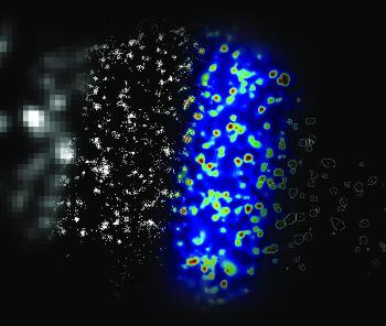 A single activated T cell
The recent findings challenge existing presumptions and identify the exact molecular switch that activates the T-cells, which could pave the way for a range of treatments in cancer and auto-immune diseases. The discovery was reported in the Nature Immunology journal by the University of New South Wales (UNSW) scientists.
The research was conducted at the Lowy Cancer Research Centre under the supervision of Associate Professor at UNSW's Centre for Vascular Research Katharina Gaus. The researchers studied a cell protein essential in early immune response and produced images of the protein molecule-by-molecule to discover the immunity 'switch' with a fluorescence microscope that was capable of achieving super-resolution.
Medical scientists could see molecules at a very high resolution of the order of 10nm with the new microscope. Dr Gaus stated that the research explained how the immune system could respond so quickly. David Williamson, a PhD candidate, whose research is based on the findings in the paper, stated that in traditional microscopy, target molecules are lit up simultaneously and individual molecules cannot be easily distinguished from the neighboring molecules.
He added that the new microscope enables target molecules to light up individually and their exact location can be determined accurately as the neighbor molecules stay dark. He also mentioned that the lighting up of the all the target molecules will enable scientists to view the sample at a 'super resolution'.
The next big discovery will be to locate other important proteins to get an overall view of T-cell activity and to improve the microscope capability to produce 3-D images at high resolution.