A pH-sensitive coating, manganese-driven chemistry, and ultrasound activation give one nanoplatform several ways to detect and attack tumors, but its performance depends on how those functions work together inside the tumor microenvironment.
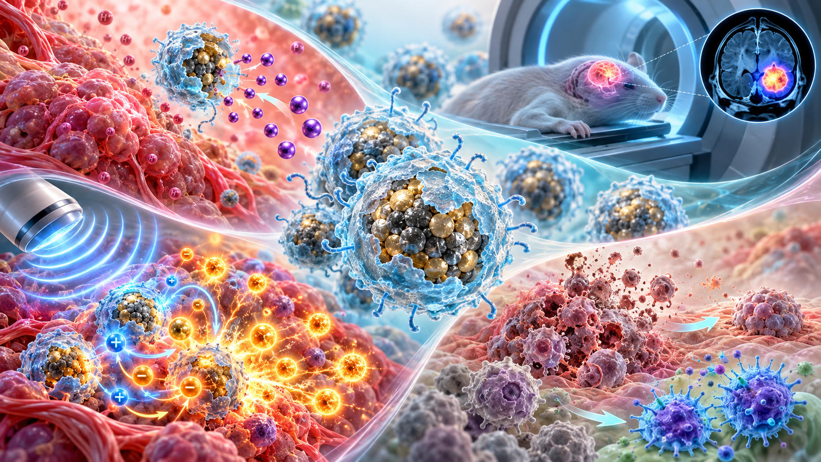
Paper: Tumor microenvironment responsive biocompatible piezoelectric nanoplatform for pH-responsive MRI guided chemo/piezodynamic therapy of subcutaneous glioma. AI-generated abstract conceptual image created using ChatGPT/OpenAI
*Important notice: This news reports on an unedited version of an accepted paper and is awaiting final editing. Therefore, the paper should not be regarded as conclusive or treated as established information.
In a recent research article published in the Journal of Nanobiotechnology, researchers developed a biocompatible, tumor microenvironment-responsive piezoelectric nanoplatform (CMBFO) for pH-responsive MRI-monitored chemodynamic/piezodynamic therapy of subcutaneous tumors derived from U87 glioma cells.
Glioma Therapy Challenges Addressed
Malignant tumors, particularly gliomas, are highly aggressive and difficult to treat because they can resist existing therapies. One possible approach is to combine piezodynamic and chemodynamic therapies so that cancer cells face both ultrasound-triggered and chemically sustained oxidative damage.
This research focuses on a cancer imaging-and-treatment platform in which acidic tumor-like conditions alter a chitosan coating and promote Mn2+ release.
The study introduces chitosan-functionalized Mn-doped bismuth ferrite (CMBFO) nanoparticles for tumor microenvironment (TME)-responsive magnetic resonance imaging (MRI) monitoring. The platform also combines chemodynamic therapy (CDT) and piezodynamic therapy (PZDT) in a subcutaneous U87 tumor model.
The researchers tested both the nanoplatform's imaging and therapeutic functions. Its design couples MRI-based tumor imaging with two complementary mechanisms that produce reactive oxygen species (ROS), which can damage tumor cells.
CMBFO Nanoplatform Design, Synthesis
The researchers designed chitosan-functionalized Mn-doped BiFeO3 (CMBFO) nanoparticles as a T2 MRI-monitored CDT/PZDT platform for tumor therapy. Bismuth ferrite (BFO) was selected as a strong piezoelectric material that can separate electrical charges under ultrasound (US) stimulation and generate ROS for PZDT.
Adding Mn2+ to the BFO lattice increased the measured piezoelectric coefficient and provided MRI contrast and catalytic activity for CDT. To improve biocompatibility and colloidal stability, the Mn-doped BFO (MBFO) nanoparticles were coated with chitosan via a sol-gel route.
The chitosan coating improved colloidal stability and provided pH responsiveness. In an in vitro blood-brain barrier (BBB) model, CMBFO showed a 38% transport ratio, higher than MBFO. Under acidic TME conditions, protonation of chitosan's amino (NH2) groups loosened the polymeric coating.
This loosening allowed Mn2+ release, activating CDT and increasing T2 MRI contrast. At neutral pH, the chitosan coating remained stable and preserved PZDT activity. The design links therapeutic activity to local pH and externally applied ultrasound.
The synthesis used defect engineering by incorporating Mn2+ into the BFO lattice to induce interstitial defects and oxygen vacancies. The authors linked these changes to improved charge separation and stronger piezoelectric and piezocatalytic responses. Controlled growth during the solvothermal process produced primary MBFO particles of 6 to 10 nm.
After chitosan coating, CMBFO formed clusters about 30 to 40 nm by transmission electron microscopy (TEM), with hydrodynamic diameters around 40 to 80 nm.
Dual ROS Activity Against Tumors
Zeta potential measurements showed pH-dependent surface charge across pH 4 to 10.
pH-tunable MRI experiments showed the strongest T2 MRI response at pH 5.1. The authors attributed this result to protonation of chitosan's -NH2 groups, which produced a positively charged, hydrophilic surface that stabilized the coating and improved interactions with water molecules.
At pH 4.0, increased protonation weakened interactions that help maintain the chitosan coating, causing the particles to aggregate more readily and reducing the MRI response. At pH 10, deprotonation lowered surface charge and hydrophilicity, weakened interactions with surrounding water molecules, and promoted agglomeration.
CMBFO also showed a stronger T2 MRI response in the presence of H2O2, which mimics part of the TME. The paper links this effect to Mn2+/Mn3+ redox cycling during a Fenton-like reaction. In mice, CMBFO accumulated preferentially in tumor tissue, and the MRI signal peaked about 6 hours after injection, with the signal-to-noise ratio rising by about 60%.
These findings were consistent with passive tumor accumulation through the EPR effect and with sustained Mn2+ release under acidic conditions. MRI measurements identified a peak in tumor signal at roughly 6 hours. Ultrasound applied at that time triggered PZDT, while the acidic TME supported sustained CDT through Mn2+ release.
The dual therapy reduced tumor volume by about 95% within 12 days. The paper reports >80% survival at day 30 in the five-mouse treatment group. Mechanistic experiments found that US-triggered PZDT produced rapid ROS bursts, while Mn-mediated CDT maintained oxidative stress and mitochondrial dysfunction. These effects were associated with apoptosis and tumor suppression.
Tumor tissue also showed a higher M1/M2 macrophage ratio after CMBFO plus US, suggesting that altered macrophage polarization may have contributed to the treatment effect. In cell experiments, CMBFO plus ultrasound caused more than 95% U87 cell death, while RAW264.7 macrophage viability remained above 95% at the tested concentrations.
CMBFO: Preclinical Findings and Remaining Questions
The study developed CMBFO as a biocompatible, multifunctional nanoplatform for MRI-monitored CDT and PZDT against subcutaneous tumors derived from U87 glioma cells. The pH-responsive chitosan coating linked MRI behavior and Mn2+ release to acidic conditions while retaining ultrasound-activated piezodynamic activity.
Rapid PZDT under ultrasound and sustained CDT through controlled Mn2+ release were associated with marked tumor suppression during a 12-day tumor-volume study, while survival was followed for 30 days. The experiments did not directly test tumor recurrence. Short-term organ histology and blood markers showed no obvious systemic toxicity within the measured endpoints.
The platform pairs piezoelectric materials, biocompatible polymers, pH-responsive imaging, and two ROS-producing treatment mechanisms.
The findings remain preclinical. Efficacy was tested in subcutaneous, not intracranial, tumors, and BBB transport was measured in vitro. Larger animal studies, pharmacokinetic work, longer-term safety testing, and studies in intracranial glioma models would be needed before the platform's relevance to patients can be assessed.
Because the tumors were outside the brain, the model does not reproduce the anatomical setting or in vivo BBB challenges of an intracranial glioma.
Source:
- Gong T., Ullah Z., et al. (2026). Tumor microenvironment responsive biocompatible piezoelectric nanoplatform for pH-responsive MRI guided chemo/piezodynamic therapy of subcutaneous glioma. Journal of Nanobiotechnology. DOI: 10.1186/s12951-026-05120-8, https://link.springer.com/article/10.1186/s12951-026-05120-8