Raman spectroscopy is a highly sensitive approach for evaluating the electrical, phonon and optical characteristics of sp2 carbon materials. Raman spectroscopy's nondestructive nature, along with both spectrum and spatial resolution, makes it an attractive contender as a standard characterization method in the rapidly expanding field of graphene.
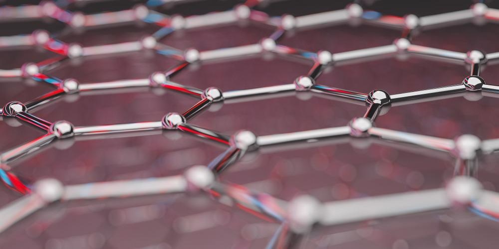
Image Credit: Production Perig/Shutterstock.com
Graphene: The Wonder Material of the 21st Century
Graphene is a two-dimensional, one-atom-thick hexagonally organized material composed of sp2 carbon networks with strong covalent bonds. As graphene absorbs only 2.3 percent of incident light, it is barely visible on most substrates. However, identifying the number of graphene layers as well as assessing the impact of disorder on its properties is crucial for the study of graphene-based electronics.
Raman Spectroscopy: Basic Overview
Raman spectroscopy is the study of the inelastic scattering of light, which is commonly related with the absorption (anti-Stokes process) and emission (Stokes process) of phonons.
Knowing the energy shift of the scattered light compared to the incident light, which generates Raman spectra, allows us to calculate the phonon frequency, which is important to convey structural information.
Raman Feature of Multilayer Graphene
In graphene, the sp2 carbon bonds result in highly polarizable π bonds that produce an intense Raman signal. The Raman spectra of graphene contain three prominent peaks of interest: the 2D peak, the G-peak, and the D-peak.
The 2D-peak (also known as the G’ peak) originates from the double resonance enhanced two-phonon lateral vibrational and this feature appears at approximately 2700 cm-1. The 2D peak becomes broader and blue-shifted when the graphene thickness increases from single layer graphene (SLG) to multilayer graphene (MLG).
The G-peak is a sharp band at roughly 1585 cm-1 that represents the in-plane stretching vibrations of the sp2 bonded carbon atoms. With increasing layer thickness, the G peak intensity increases linearly until 10 layers (3 nm) and then drops with increasing sheet thickness.
The number of graphene layers can be determined using the intensity ratio (I2D/IG) and the FWHM of the 2D peak. For example, with high-quality SLG, the I2D/IG ratio will be 2.
The D peak is commonly referred to as the disorder band. This peak is caused by lattice motion away from the Brillouin zone's centre, occurring between 1270 and 1450 cm-1. Graphene-based materials having defects, such as graphene oxide, disordered graphene, and nanographene, can also be identified using the D peak.
Raman Spectroscopy of Pristine Single Layer Graphene
SLG has two atoms per unit cell, resulting in six normal modes at the center of Brillouin zone, two of which are doubly degenerate: A2u + B2g + E1u + E2g. Aside from the G mode, the Raman spectra of pristine SLG consist of 2D and 2D’ peaks that occur around 2700 and 3240 cm-1, respectively.
The D and D' peaks, which are the fundamental modes of the 2D and 2D' peaks, need a defect to be activated in the double resonance Raman scattering process (DRRS), and so are missing from the Raman spectra of pristine SLG.
Edges are a type of defect that occur naturally in every graphene sample because of the broken translational symmetry. As a result, at SLG's edge, the D and D′ peaks can be seen in the Raman spectrum.
Recent Research on Graphene Raman Spectroscopy
The electrical and optical properties of multilayer graphene are significantly influenced by the stacking order. As a result, Raman spectroscopy may be used to investigate MLG stacking order. For example, when comparing ABC-stacked 3LG (1581 cm-1) to AB-stacked 3LG (1582 cm-1), the G mode of ABC-stacked 3LG (1581 cm-1) is 1 cm-1 red shifted. Also, the 2D mode in ABC-3LG, is more asymmetrical.
Furthermore, graphene's electrical and lattice vibration properties alter dramatically in the presence of external perturbations such as doping, defects, stress, strain, temperature, and magnetic fields, which can be evaluated via Raman spectroscopy.
The G peak, for example, sharpens with both hole and electron doping, while the 2D peak blueshifts monotonically when p-doped but redshifts when n-doped. The intensity ratio of I(2D)/I(G) can be used to determine the level of doping.
Applying external strain on graphene can potentially open the bandgap. The G mode splits into two peaks, G+ and G−, when a uniaxial tension strain is applied.
The FWHM for the 2D peak remains unchanged, but the peak can redshift and split due to the changing band structure. In addition, Raman spectroscopy can be utilized to detect structural degradation, undesirable by-products, functional groups, and chemical alterations introduced during graphene synthesis.
Raman Mapping of Graphene Layers
Graphene is notoriously difficult to locate optically; nevertheless, Raman mapping can easily visualize the distribution of 1–4 LG. In addition, Raman imaging clearly shows the defect band at some edges.
Furthermore, the isotope labeling Raman imaging approach may also be used to assess graphene growth CVD parameters, such as methane gas flow rate and temperature, and is commonly employed to uncover the graphene growth mechanism.
Conclusion
Raman spectroscopy is an integral part of studying graphene-based materials such as graphene quantum dots, graphene nanoribbons, graphene composites and many more.
Raman spectroscopy and imaging have the advantage of not being dependent on the substrate. This is due to the fact that the Raman spectrum is an intrinsic property of graphene. Conclusively, Raman spectroscopy has been demonstrated to be a nondestructive and real-time approach for monitoring graphene-based devices.
Continue reading: What is Twisted Graphene?
References and Further Reading
Wu, J., Lin, M., Cong, X., Liu, H. and Tan, P., (2018) Raman spectroscopy of graphene-based materials and its applications in related devices. Chemical Society Reviews, 47(5), pp.1822-1873. Available at: https://doi.org/10.1039/C6CS00915H
Disclaimer: The views expressed here are those of the author expressed in their private capacity and do not necessarily represent the views of AZoM.com Limited T/A AZoNetwork the owner and operator of this website. This disclaimer forms part of the Terms and conditions of use of this website.