Microscopy, spectroscopy, and atomistic modeling trace rare earths through an unexpected nanoscale mineral environment in Pacific Ocean sediments.
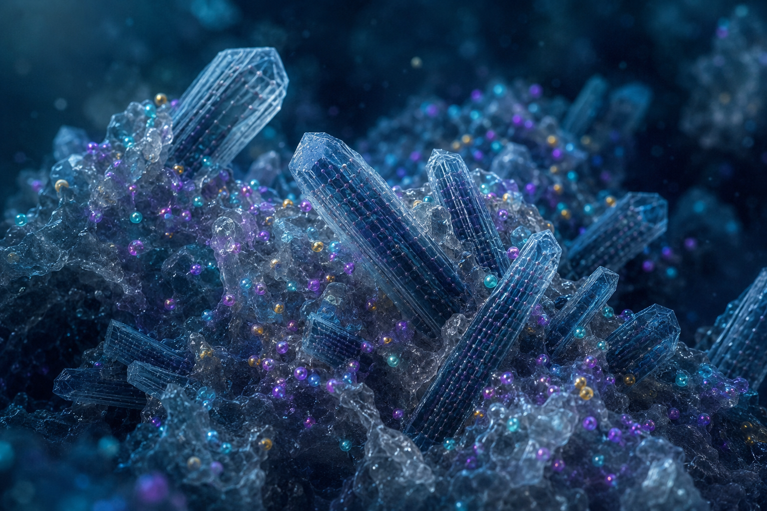
Paper: The atomic-scale structure of rare earth elements in deep-sea sediments facilitates their extractability. Image credit: AI-generated image created using ChatGPT/OpenAI
In a recent research article accepted for publication in the journal Communications Earth & Environment, researchers investigated the nanocrystalline architecture of carbonate fluorapatite and the atomic-scale environment of rare-earth elements (REEs) in deep-sea sediments to elucidate their unexpectedly high extractability despite fluorapatite's low solubility.
REEs in Marine CFA
Rare earth elements (REEs) are critical materials widely used in many advanced technologies and green energy applications. Terrestrial sources of REEs are increasingly limited, driving interest in alternative reservoirs such as marine sediments.
Deep-sea sediments have been identified as a promising source of REEs, with these elements commonly found in carbonate fluorapatite (CFA), a mineral comprising calcium, phosphate, carbonate, and fluoride. Two forms of CFA are present in such sediments: biogenic CFA (b-CFA), derived from the remains of marine organisms, and authigenic CFA (a-CFA), formed in situ during sediment burial.
Although fluorapatite (FAp) has low solubility, acid leaching readily recovers REEs from pelagic sediments, raising questions about their exact structural incorporation. Understanding the atomic-scale location and bonding environment of REEs in CFA could help explain their extractability and inform the development of more efficient and potentially safer recovery strategies.
Nanostructure and Spectroscopy Analysis
The study investigated the nanostructure and atomic arrangement of samarium (Sm), a proxy for REEs, within biogenic and authigenic CFA grains collected from Pacific Ocean sediment cores. High-resolution transmission electron microscopy (HRTEM) and fast Fourier transforms (FFT) were used to characterize the morphology and crystallinity of the CFA nanograins.
Extended X-ray absorption fine structure (EXAFS) spectroscopy, measured in high-energy-resolution fluorescence detection (HERFD) mode at cryogenic temperatures, provided enhanced structural resolution and signal-to-noise ratio for the local atomic environment of Sm.
Comparative EXAFS analyses employed references including magmatic fluorapatite (m-FAp), in which Sm is lattice-incorporated, and Sm-adsorbed hydroxyapatite (HAp), in which Sm is surface-bound. Density functional theory (DFT) calculations assessed Sm-REY pairing in apatite and compared the affinities of Sm and cerium (Ce) for apatite- and monazite-type environments, providing atomistic insights.
Additional thermodynamic modeling addressed the solubility behavior of REY (rare earth elements and yttrium) phosphates in seawater. The study also qualitatively examined CFA dissolution following acid leaching by imaging residual particles with TEM and analyzing them using energy-dispersive X-ray spectroscopy (EDS).
Samarium Localization and Extractability in Nanocrystalline CFA
At the nanoscale, neither a-CFA nor b-CFA is a uniform crystal; instead, both are composite materials composed of poorly crystalline apatite nanorods embedded within an amorphous matrix. The nanorods were elongated primarily along the [001] crystallographic direction, with a-CFA rods generally longer than those in b-CFA, a difference consistent with their distinct formation processes. This composite nanoarchitecture helps explain REY recovery because both the amorphous host matrix and CFA nanocrystals dissolve more readily under acid leaching than larger, well-crystallized fluorapatite.
EXAFS analyses showed that Sm’s local structural environment in both a-CFA and b-CFA is similar and distinct from those of the reference compounds. In magmatic fluorapatite, Sm is incorporated into a crystalline lattice site, coordinated to oxygen, phosphorus, and calcium atoms in a well-ordered arrangement.
Conversely, in CFA sediments, Sm resides predominantly in an amorphous phase surrounding the apatite nanocrystals rather than substituting directly for calcium within the apatite lattice. Sm is coordinated by approximately eight oxygen atoms and has more distant phosphorus and calcium neighbors in apatite-like linkages, but its local environment exhibits a high degree of positional disorder, as evidenced by the lack of resolved atomic pairs beyond ~5 Å in radial structure functions.
DFT modeling suggested that Sm preferentially forms pairs with other REY atoms at medium-range distances (~6.2–6.3 Å), while calcium remains favored at shorter distances (~4.0–4.2 Å), a result consistent with the EXAFS findings.
The calculations also indicated that Sm favors an apatite-type bonding environment, whereas cerium exhibits a stronger affinity for Ce-phosphate bonding environments akin to those in less soluble monazite minerals. Thermodynamic solubility modeling predicted that Ce phosphate could precipitate under simulated seafloor seawater conditions, which may help explain Ce's lower extractability compared to other REYs. The modeling also predicted Y-phosphate saturation across part of the examined pH range.
A qualitative acid-leaching experiment examined the dissolution of CFA under acidic conditions. A brief exposure (3 minutes) of CFA particles to 0.25 M hydrochloric acid almost completely dissolved the biogenic apatite teeth and significantly reduced calcium and phosphate signals in residual particles, which mainly comprised micaceous silicate phases. These findings are consistent with the faster dissolution of the amorphous calcium phosphate matrix compared to the remaining nanocrystalline CFA. No REYs were clearly detected in the residues, and the experiment did not quantify REY recovery efficiency.
The nanocrystalline component of CFA is also more soluble than macro- and microcrystalline fluorapatite, facilitating REY recovery. The lower leachability of Ce is consistent with its incorporation within less-soluble Ce-phosphate phases, precipitation as CeO2, or incorporation into ferromanganese nodules.
Implications for REE Recovery
This study highlights the critical role of the atomic-scale structure of REYs in deep-sea sediments in governing how readily they can be extracted. Samarium is primarily bound within an amorphous apatitic matrix surrounding CFA nanocrystals rather than substituting directly into the apatite lattice.
Understanding these local bonding environments could inform the development of more efficient and potentially safer REY extraction methods from marine deposits.
This insight into the nanoscale mineralogy and bonding of critical elements shows how nanocrystal dimensions, crystallinity, and surrounding amorphous phases can influence the accessibility of strategically important elements and may guide future resource recovery strategies.
Source:
- Manceau, A., Giacomelli, A., Li, Y., Gaillot, A. C., Liao, J., Spadini, L., Koschinsky, A., Mathon, O., & Steinmann, S. N. (2026). The atomic-scale structure of rare earth elements in deep-sea sediments facilitates their extractability. Communications Earth & Environment. DOI: 10.1038/s43247-026-03848-7, https://www.nature.com/articles/s43247-026-03848-7