A research team from the Brown University has developed a first-of-its-kind three-dimensional live tissue model of a glioma or brain tumor surrounded with a network of blood vessels to assess the efficacy of brain cancer drugs. The research findings have been reported in the journal Theranostics.
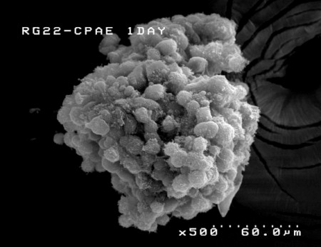 The 3-D glioma model, developed at Brown University, allows the glioma and the supporting endothelial cells to assemble naturally, just as they would in real life. (Credit: Sun Lab/Brown University)
The 3-D tissue model is a concept of Jeffrey Morgan, who is a bioengineer at the Brown University. Lead author Don Ho and other researchers formed an agarose hydrogel mold wherein a rat RG2-cell glioma with a diameter of 200 µm was created. They then enclosed the tumor with endothelial cells obtained from cow respiratory vessels to form the network of blood vessels.
In the 3-D model, the endothelial cells are attached with the tumor instead of being isolated from the substrate as in the case of Petri-dish-type analyses. This significant advantage allows scientists to probe the formation and growth of endothelial cells and the action of anti-therapeutic agents, as they are in a live organism.
The researchers then dosed the mold after attaching the tumstatin, an element of a natural protein present in collagen, with iron-oxide nanoparticles. As expected, the endothelial cells gobbled up the nanoparticles. In a string of in vitro tests, the researchers discovered that the tumstatin iron-oxide nanoparticles achieved a 2.7 times more reduction in vasculature growth than under standard conditions over 8 days. The growth of new endothelial cells was completely blocked.
The use of iron-oxide nanoparticles is highly important, as they are readily gobbled up by endothelial cells and easily traced by magnetic resonance imaging. The 3-D tissue model clearly demonstrated the ability of the iron-oxide nanoparticles to reach blood vessels enclosing the glioma and also the ability of tumstatin to pierce endothelial cells.
Ho explained that this model paves way to test and optimize the design of diagnostic/therapeutic nanocarriers and detect their therapeutic capabilities. It may help in saving money and time, while decreasing tests in live organisms to assess the 3-D characteristics of an agent such as its capability for diffusion and targeting. The researchers’ next step is to assess the performance of the tumstatin nanoparticles in the 3-D environment, Ho concluded.