The HT7800 series comes in three variants: The HT7800, HT7820, and HT7830 (see Specifications table below). These microscopes represent a new era of transmission electron microscopy, combining Hitachi's 75+ years of expertise with modern digital advancements. Designed for both seasoned researchers and newcomers, this series offers high-resolution imaging with a user-friendly interface and ergonomic controls that enhance efficiency and comfort.
Designed for life sciences, material sciences, and industrial applications, the HT7800 series provides flexible imaging modes, high-speed digital processing, and seamless automation, ensuring accurate and reproducible results. You can also operate the microscope in normal room light conditions, which removes the need for a darkroom and improves usability and comfort.
- Dual-Mode objective lens: Switch between high-contrast and high-resolution modes with a single click for unmatched flexibility.
- User-friendly interface: Modern, intuitive GUI enables seamless operation under normal room lighting conditions.
- Enhanced digital imaging system: Includes low-dose functionality to protect sample integrity.
- Expandable capabilities: Compatible with STEM, EDX, MirrorCLEM, and other specialized accessories to meet diverse research needs.
Features and Benefits
Dual-Mode Objective Lens for Maximum Versatility
- Easily toggle between high-contrast and high-resolution modes on a single microscope
- Removes the need for multiple instruments, streamlining workflows and reducing costs
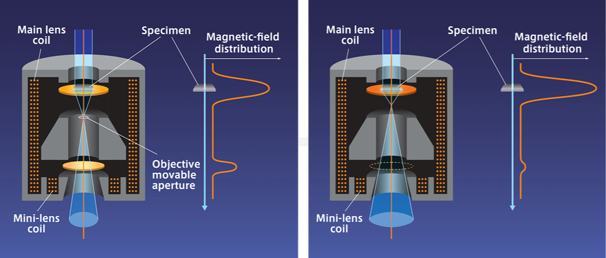
. Image Credit: Hitachi High-Tech Europe
Intuitive Operation and User-Friendly Interface
- Designed for both novice and expert users, the HT7800 series features a streamlined digital GUI.
- With its integrated CMOS screen camera, the system can be operated in a well-lit room, reducing fatigue and enhancing ergonomics.
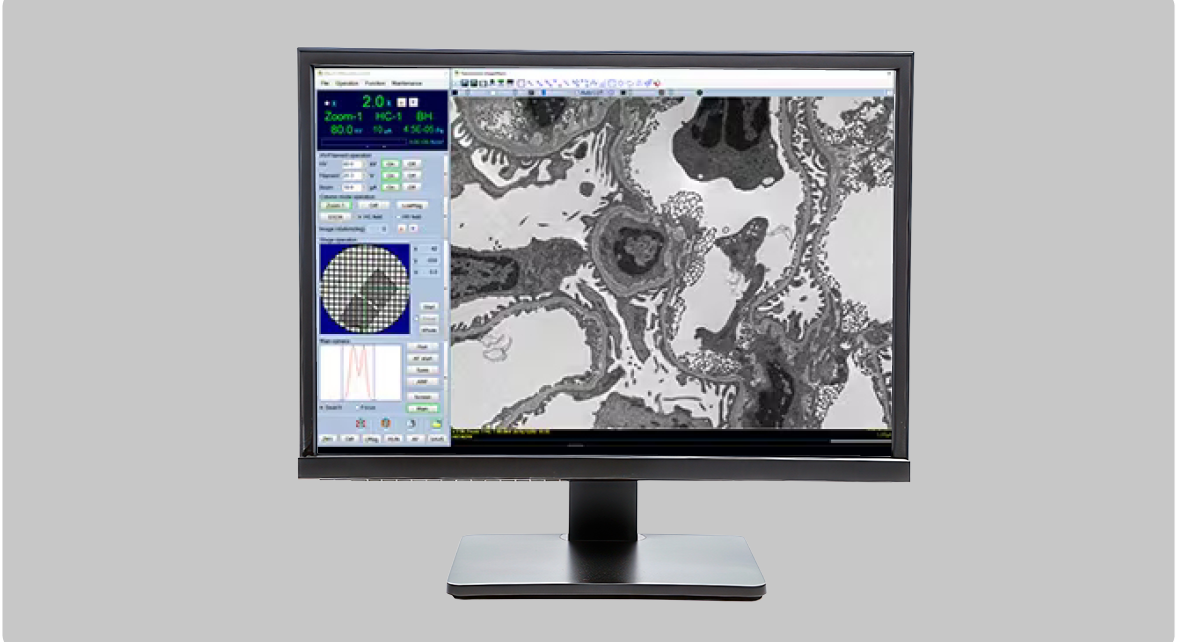
. Image Credit: Hitachi High-Tech Europe
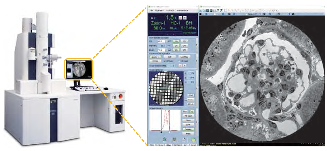
The HT7800’s Digital Imaging System
- A high-speed CMOS camera for superior imaging
- The Whole View Function enables fast, automated image acquisition over large areas
. Image Credit: Hitachi High-Tech Europe
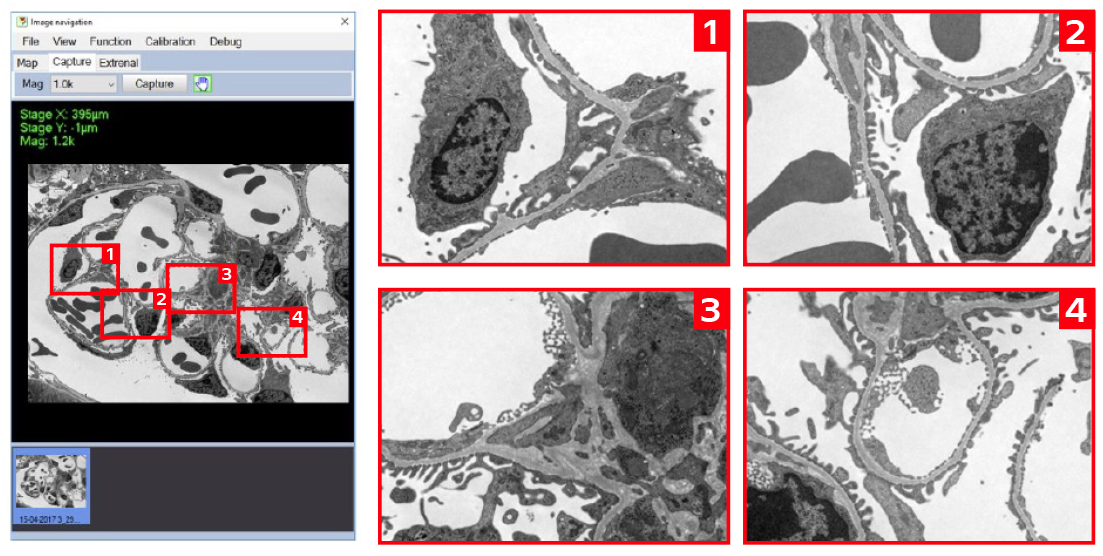
Automation and Navigation
- Auto Multiple Frame (AMF) Imaging: Quickly stitch multiple images together to create high-resolution panoramic images.
- 3D Electron Tomography: It captures tilted images for accurate 3D reconstructions.
- Advanced Image Navigation and Mapping: Quickly pinpoint and analyze the regions of interest.
. Image Credit: Hitachi High-Tech Europe
Expandable and Customizable for Advanced Research Needs
- STEM and EDX: Enables detailed elemental analysis and material characterization.
- MirrorCLEM: Correlates fluorescence microscopy with TEM imaging, providing more comprehensive research insights.
. Image Credit: Hitachi High-Tech Europe
Specifications
Source: Hitachi High-Tech Europe
| |
HT7800 |
HT7820 |
HT7830 |
| Electron gun |
W (standard), LaB6 |
LaB6 (standard), W |
LaB6 (standard), W |
| Accelerating voltage |
20 - 120 kV (100 V/step variable) |
20 - 120 kV (100 V/step variable) |
20 - 120 kV (100 V/step variable) |
| Resolution (Lattice) |
0.20 nm (Off-axis, 100 kV) |
0.14 nm (Off-axis, 120 kV) |
0.14 nm (Off-axis, 120 kV)
0.19 nm (On-axis, 120 kV) |
| Maximum magnification |
x600,000 |
x800,000 |
x1,000,000 |
| Stage maximum tilt angle |
±70° |
±30° |
±10° |
Standard
features |
Auto focus, Microtrace, Autodrive, Live FFT display, Measurement function, Low dose, API (auto pre-irradiation),
Image navigation function, Column with mild baking function, Whole view function, Drift correction function, etc. |
Applications Gallery
Life Sciences and Biomedical Research
- High-contrast imaging for biological specimens
- Low-dose mode for cryo-TEM and sensitive biological samples
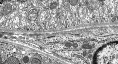
The images below illustrate a comparison between a conventional stained section and an unstained section using the HT7800 series High-Contrast lens.
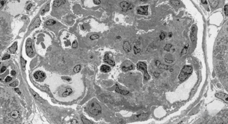
Mouse Kidney (stained), accelerating voltage 80 kV, magnification x300. Image Credit: Hitachi High-Tech Europe
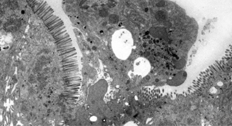
Rat jejunum (unstained), accelerating voltage 80 kV, magnification x2,000. Image Credit: Hitachi High-Tech Europe
Material Science and Nanotechnology
- High-resolution imaging of nanoparticles, polymers, and advanced materials
- STEM & EDX compatibility for detailed compositional analysis
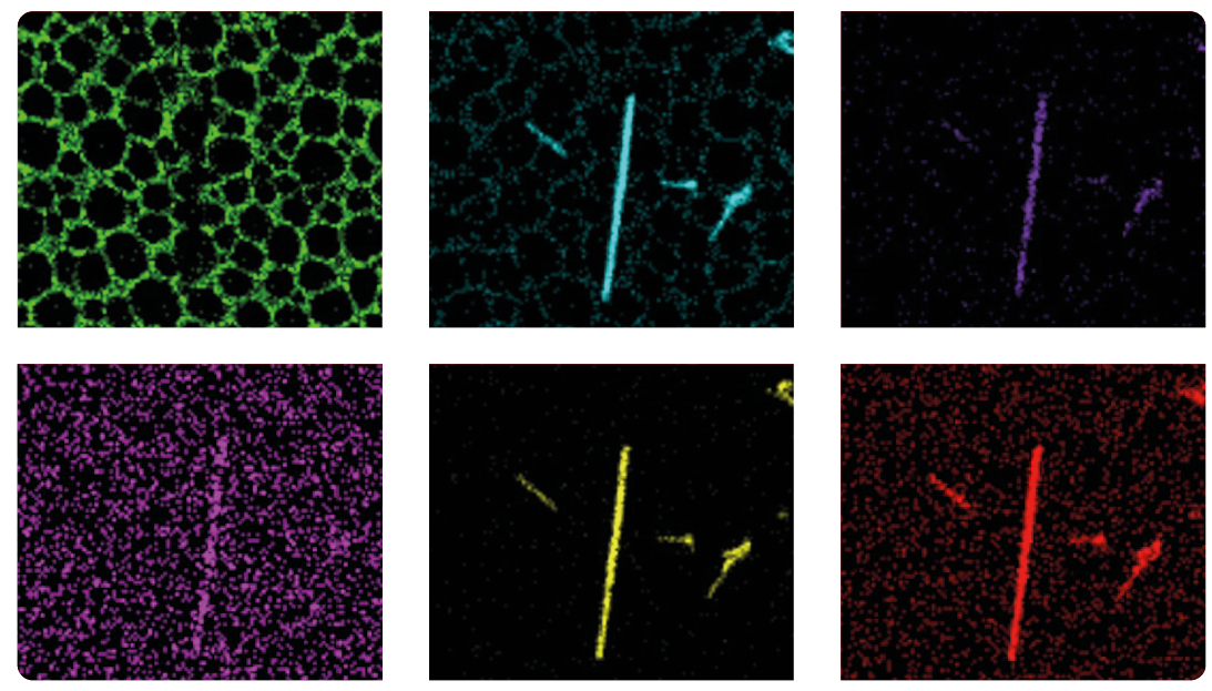
The image below shows hollow-cone dark-field observation of an Au/TiO2 catalyst on the HT7820. The electron beam diffraction region used for the hollow-cone dark-field observations is highlighted in yellow within the selected-area electron diffraction pattern. In the resulting images, TiO2 diffraction waves are clearly visible.
Au/TiO2 catalyst. Bright-field TEM image, accelerating voltage 120 kV. Image Credit: Hitachi High-Tech Europe
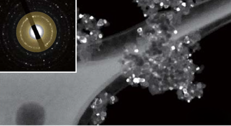
Au/TiO2 catalyst. Hollow-cone dark-field TEM image, accelerating voltage 120 kV. Image Credit: Hitachi High-Tech Europe
Particle/Polymer
- High-resolution imaging and digitization for rapid observation of sequences of nanometer-scale pores
The images below show TEM results of mesoporous silica particles, intended for application in drug delivery systems.
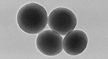
Mesoporous silica powder, accelerating voltage 120 kV, magnification x70,000. Image Credit: Hitachi High-Tech Europe
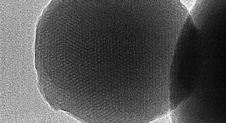
Mesoporous silica powder, accelerating voltage 120 kV, magnification x200,000. Image Credit: Hitachi High-Tech Europe