Due to its impressive ability to discern nanoparticles based on their shape, size and environment, hyperspectral darkfield microscopy is rapidly being adopted by several scientific communities. Nanoparticles toxicology, immunology, microbiology and advanced material are all benefiting from this new capacity to locate and identify nanoparticles.
The smaller the object, the dimmer it is and nanoparticles are no different. This is why hyperspectral darkfield microscopes require intense illumination in order to produce high quality images. However, an intense and prolonged illumination can damage tissues and living organisms,forcing careful researchers to tediously manage light exposure. Inorganic nanoparticles may also degrade by absorbing the heat generated by intense and prolonged white light exposure.
To circumvent this limitation, Photon etc has produced a novel hyperspectral darkfield microscope based on a tunable light source that selectively delivers monochromatic light, decreasing heat transfer to the samples by two orders of magnitude compared to white light illumination. This hyperspectral darkfield microscope takes advantage of Photon etc.’s patented filtering technology and illuminates the entire field of view of a research-grade microscope with a continuously tunable, bright laser light.
To produce hyperspectral data, darkfield microscopy images are captured while the tunable light source’s emission is quickly swept across its emission range (400 to 1000 nm or 400 to 1650 nm).
This instrument also offers new possibilities to researchers. Since the tunable source can rapidly switch between selected wavelengths, the user can select those resonant with specific nanoparticles and track their movement in real time across the hyperspectral microscope’s field of view. For example, this feature could be beneficial to study the dynamics of nanoparticle drug delivery in cancerous cells and other in-vitro samples.
LIMA allows the imaging of particlesof a few nanometers in diameter. Detecting these very small particles is of great importance, as they can freely penetrate cellular membranes and cell nuclei.
While this instrument is primarily dedicated to darkfield microscopy, it can be easily converted into a brightfield hyperspectral imaging microscope by simply interchanging the darkfield condenser by a brightfield condenser.
This hyperspectral darkfield microscope provides a high spectral resolution in the VIS, NIR, and SWIR ranges, combined to a near-diffraction-limited spatial resolution. Ideal for darkfield microscopy, PLE, or standard brightfield reflectance and transmittance imaging, it can map the full spectral response of an extended range of samples.
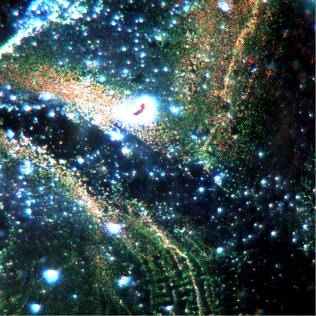
High-resolution dark-field image reveals intricate details of nanoparticles embedded in organic fibers.
Image Credit: Photo etc.
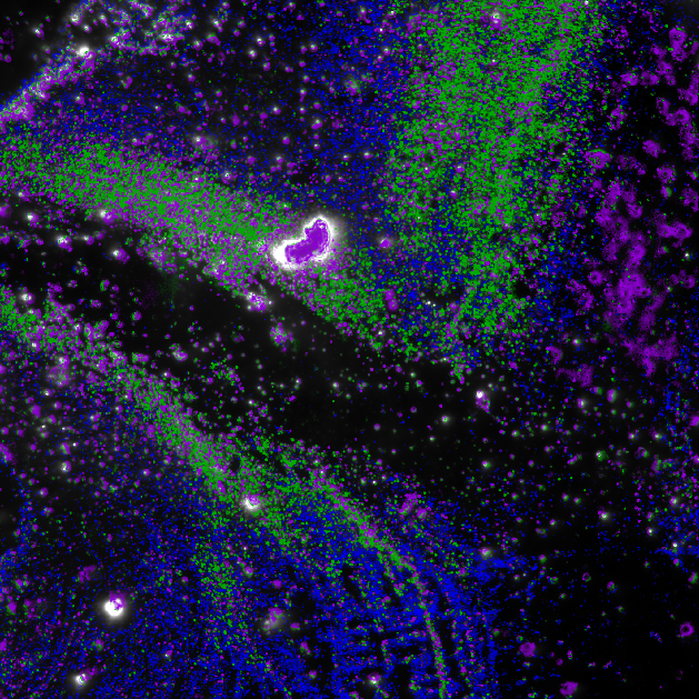
Spectral Angle Mapping (SAM) analysis unveils the primary nanoparticle families imaged by the hyperspectral dark field microscope.
Image Credit: Photo etc.
Key Features
The main features of the LIMA are:
- VIS, NIR and/or SWIR spectral ranges. One single instrument covering 400 nm - 1620 nm
- Darkfield, photoluminescence excitation, or standard brightfield reflectance and transmittance hyperspectral imaging
- Continuously tunable fluorescence excitation source
- High sensitivity/Low noise MP s-CMOS camera /InGaAs camera
- High spatial and spectral resolution
- Scientific grade microscope
- PHySpec Software: acquisition and analysis – unlimited license included
- Customization available
Applications
Nanoparticles imaging and classification
Contact Photon etc for more information on these applications
|
Technical Specifications
|
VIS-NIR |
VIS-SWIR |
| Spectral Range |
400 - 1000 nm |
400 - 1620 nm |
| Spectral Resolution |
1.5 - 2.5 nm |
< 4 nm |
| Spatial Resolution |
Sub-micron ; limited by the microscope objective NA |
| Camera |
sCMOS (EMCCD) |
sCMOS (EMCCD) + Photon etc. InGaAs camera (ZephIR 1.7 or Alizé 1.7) |
| Excitation Wavelength |
Continuously tunable |
| Microscope |
Upright or inverted, scientific grade |
| Illumination |
High efficiency homogeneous illumination; Dia or Epi illumination; Brightfield; Darkfield (oil and dry) |
| Visualization mode |
Hyperspectral, and broadband visualization modes |
| Preprocessing |
Spatial filtering, statistical tools, spectrum extraction, data normalization, spectral calibration |
| Hyperspectral Data Format |
FITS, HDF5 |
| Software |
Computer with PHySpec™ control and analysis software included |
| Dimensions |
≈ 105 cm x 82 cm x 82 cm |
| Weight |
≈ 80 Kg |
| Options |
| Spectral range |
1000-2500 nm in one instrument
UV option |
| XY motorized stage |
100 mm x 100 mm travel, 22 mm resolution |
LIMA - Hyperspectral Scanning Excitation Darkfield Microscope